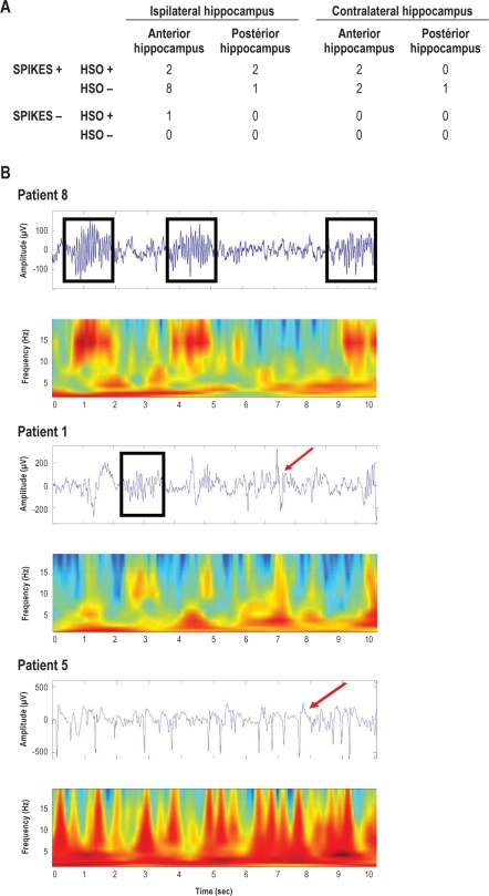
Figure 5.
Hippocampal spindle frequency oscillations (HSO). The table shows the distribution (number of patients) of HSO according to the presence of spikes and the location. No significant correlation was found for presence vs absence of HSO according to the side (ipsilateral to the epileptic focus vs contralateral), the site (anterior vs posterior hippocampus), and the presence or the absence of spikes (χ2, P > 0.3, n = 21). Examples of hippocampus EEG traces and time-frequency analysis (10 sec) during SWS sleep (spikes = red arrow, spindles = black squares). Top: no spikes and HSO (Patient 8); Middle: spikes and HSO (Patient 1); Bottom: spikes and no HSO (Patient 5).

