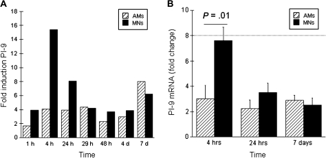
Figure 1.
Differing kinetics of Mycobacterium tuberculosis–induced serine protease inhibitor 9 (PI-9) expression in alveolar macrophages (AMs) and blood monocytes (MNs). A, Representative study of the kinetics of PI-9 expression as assessed from 1 hour through 7 days after infection of AMs (striped bars) and MNs (solid bars) with M. tuberculosis strain H37Rv. Expression of PI-9 mRNA is represented as fold induction (PI-9 messenger RNA [mRNA] within M. tuberculosis infected cells as determined by real-time polymerase chain reaction divided by that of uninfected cells collected at the same time point). As illustrated, MNs displayed a greater early peak of PI-9 mRNA than did AMs (at 4 hours after M. tuberculosis infection), but PI-9 expression persisted in both cell types throughout the 7 day course of the experiment. B, Mean data from studies of 16 subjects confirmed that early differences in PI-9 expression resolved rapidly. As illustrated, H37Rv induced a 7.6-fold increase in PI-9 mRNA in MNs (solid bars) at 4 hours, compared to a 3.2 fold increase in AMs (striped bars). By 24 hours, however, PI-9 expression in MNs had declined significantly, so that induction of PI-9 was similar within M. tuberculosis–infected AMs and MNs. PI-9 expression continued through day 7 in both cell types, as illustrated.

