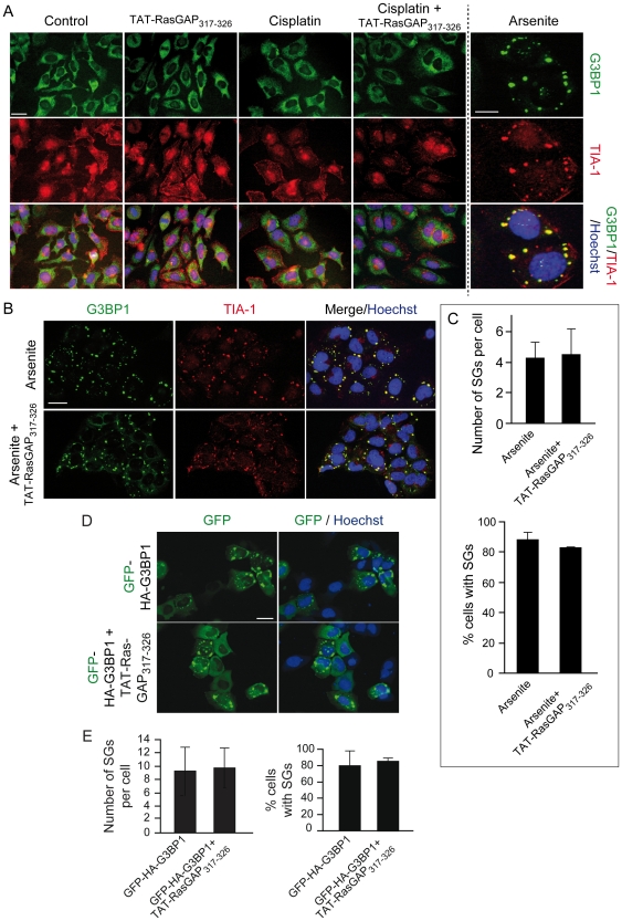
Figure 5. TAT-RasGAP317–326 does not affect G3BP1-induced SG formation.
A. U2OS cells were left untreated or incubated with 15 µM cisplatin, 20 µM TAT-RasGAP317–326, or a combination of the two compounds for 22 hours. Alternatively, the cells were treated with 200 µM arsenite for 2 hours. The cells were then processed for immunofluorescence analysis using TIA-1- and G3BP1-specific antibodies. Nuclei were stained with Hoechst 33342. Images were taken with a fluorescent microscope (Nikon Eclipse 90i). Scale bar: 20 µm for the first 4 columns, 5 µm for the last column. B–C. U2OS cells were pre-incubated or not with 20 µM TAT-RasGAP317–326 for 18 hours and then treated with arsenite (200 µM, 2 hours). The cells were then processed as in panel A. Scale bar: 20 µm. Quantitation of the number of SGs per cells and the percentage of cells with SGs is shown in panel C. D–E. U2OS cells were transfected with a plasmid encoding GFP-HA-G3BP1 and 6 hours later were incubated or not with 20 µM TAT-RasGAP317–326 for an additional 22 hour period. Cells were then fixed and their nuclei stained with Hoechst 33342. Scale bar: 20 µm. Panel E shows the quantitation of the number of SGs per cells as well as the percentage of cells with SGs.

