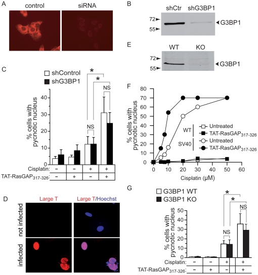
Figure 8. G3BP1 silencing does not affect TAT-RasGAP317–326-mediated sensitization of cancer cells to cisplatin-induced apoptosis.
A, B. U2OS were infected with a non-target shRNA control vector or a G3BP1 shRNA-expressing lentivirus. After 72 hours, cells were analyzed by immunocytochemistry (panel A) and Western blotting (panel B) for the presence of G3BP1. C. Infected cells were incubated with 20 µM cisplatin and 20 µM TAT-RasGAP317–326 for 22 hours as indicated on the figure. Apoptosis was then scored. D. MEFs were infected or not with a large T antigen-expressing lentivirus and 3 days later the expression of the large T antigen was assessed by immunocytochemistry. E. G3BP1 expression in wild-type (WT) and G3BP1 knock-out (KO) SV40 large T antigen-transformed MEFs was assessed by Western blotting. F. Alternatively, these cells were incubated with increasing concentrations of cisplatin in the presence or in the absence of TAT-RasGAP317–326 for 22 hours. Apoptosis was then measured by scoring the number of cells with pycnotic nuclei. G. Wild-type (WT) and G3BP1 knock-out (KO) SV40 large T-transformed MEFs were treated as in panel C. Asterisks indicate statistically significant differences; NS: not significant.

