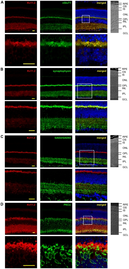
Figure 4. Double-labeling experiments for the identification of Kv11.2 channel subunit expressing cells in the mouse retina.
(A) The presynaptic marker of glutamatergic synapses vGluT1 was used for double-labeling experiments with Kv11.2. The merged data show a high degree of co-localization of both proteins. The box highlights the section shown in the bottom row in higher magnification illustrating the co-expression of Kv11.2 and vGluT1 in the IPL and OPL. (B) Double-labeling for the unspecific presynaptic marker synaptophysin and for Kv11.2. The merged data show a high degree of overlap in the OPL. In the IPL, Kv11.2 immunoreactivity is present in a subset of synaptophysin-positive presynaptic structures. The box highlights the section shown in the bottom row in higher magnification. (C) Double-labeling for the GABAergic marker GAD2/GAD65 and for Kv11.2. The merged data show no co-localization of both proteins. The box highlights the section shown in the bottom row in higher magnification illustrating the distinct expression of Kv11.2 and GAD2/GAD65. (D) A PKCα-specific antibody was used for double-labeling experiments with the Kv11.2-specific antibody. Some co-localization was detected in the IPL, especially in the axonal terminals of bipolar cells. In the OPL no co-localization could be detected. The box highlights the section shown in the bottom row in higher magnification illustrating the distinct expression pattern of Kv11.2 and PKCα in the OPL. On the right of each panel a part of the bright field picture is shown. RPE: retinal pigment epithelium; OS: outer segments; IS: inner segments; ONL: outer nuclear layer; OPL: outer plexiform layer; INL: inner nuclear layer; IPL: inner plexiform layer; GCL: ganglion cell layer. Scale bars: 20 µm. The results of counter staining of cell nuclei (blue) is included in the merged pictures shown on the right.

