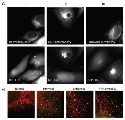
Figure 7.
Nsp6 orthologs from MHV, SARS-CoV and PRRSV induce vesicles containing LC3 and show differences in extent of incorporation into autophagosomes. (A) (i–iii) MHVnsp6mCherry, SARSnsp6mCherry and PRRSVnsp567mCherry, were expressed in CHO cells expressing GFP-LC3. The distribution of the proteins was determined from their natural fluorescence. GFP-LC3 signals are shown in the lower parts. (B) IBVnsp6mCherry, MHVnsp6mCherry, SARSnsp6mCherry and PRRSVnsp567mCherry, were expressed in CHO cells expressing GFP-LC3. The distribution of the proteins was determined from their natural fluorescence. High magnification images were merged to determine extent of colocalization between the nsp6 orthologs and GFP-LC3. Notably vesicular structures containing SARS-CoV colocalize with LC3-GFP.

