


Figure 2.
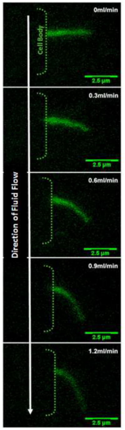
XZ bending profiles of a kidney cell primary cilium under increasing flow rates. There is a one-to-one functional relationship between flow rate and degree of bending. Cilia are visualized by transfection with a SSTR3-GFP fusion protein which specifically localizes to the cilium.