
Fig. 1.
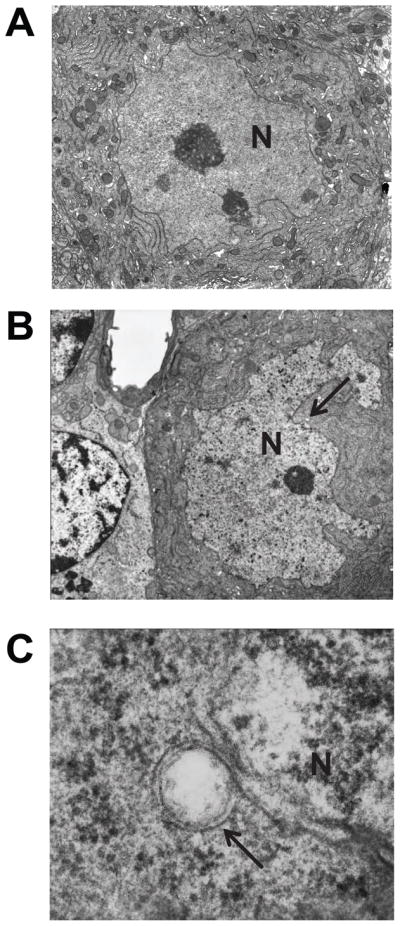
Nuclear envelope structures of the cerebellar Purkinje cells in WT and Sgce KO mice. Although no abnormal nuclei were detected in the cerebellar Purkinje cells of WT mice (A), abnormal nuclei were detected in those of Sgce KO mice (B). Enlarged and inverted images of B in Sgce KO mice (C) revealed that the nuclear envelope blebbing and other nuclear envelope abnormalities were similar to those reported in DYT1 dystonia models. Magnifications; A, B: 5,000 ×; C: 100,000 ×. Nuclei (N) are marked in the images. Arrows in B and C indicate the abnormal nuclear envelopes. Representative electron microscope images are shown.