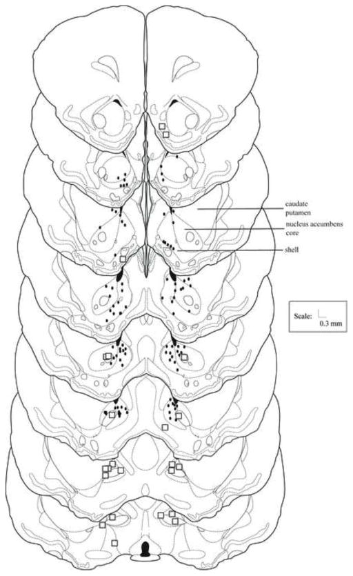
Figure 1.
Nucleus accumbens histology. Histological reconstruction of injection sites of animals receiving infusions into the nucleus accumbens shell or core 5 min before defeat (Experiment 1) or testing (Experiment 2). Black dots represent the site of injection of one or more animals. Boxes represent one or more anatomical misses. Drawings adapted from [59].

