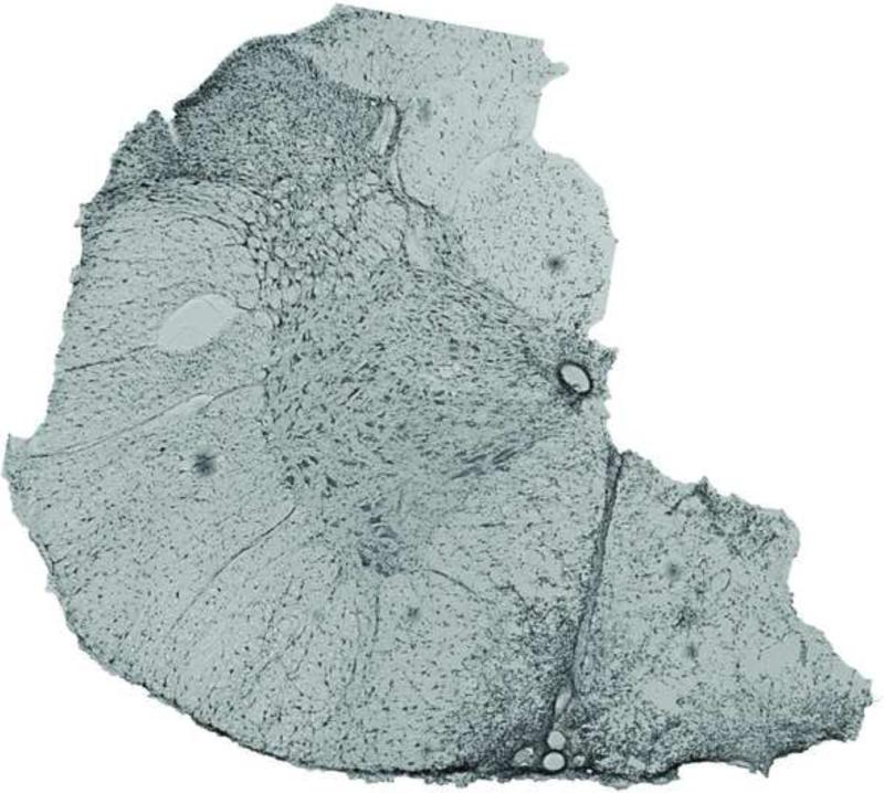
Figure 1.
A coronal section taken from the site of C2 lateral section shows a typical extent of injury. C2 lateral section was incomplete in these studies, with the ventromedial funiculus remaining intact. These images are similar in many respects to many previous studies concerning spontaneous recovery of respiratory motor function following cervical spinal hemisection (Fuller et al., 2009; Goshgarian, 1981). The micrograph shows a full coronal section and was obtained using phase-contrast microscopy.

