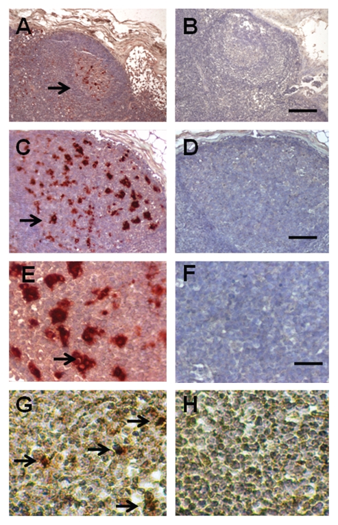
Figure 2.
Sections of a mesenteric lymph node from a macaque transplanted with E28 pig pancreatic primordia in mesentery, followed by porcine islets in the renal subcapsular space stained using anti-insulin antibodies (A, C and E) or control antiserum (B, D and F) or hybridized to an antisense (G) or sense (H) probe for porcine proinsulin mRNA. Arrows, positively staining cells (A, C, E and G). Scale bars, 80 µm (A and B); 25 µm (C and D); 15 µm (E–H).

