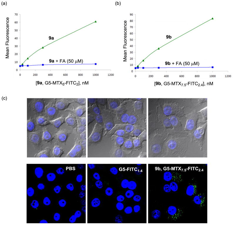
Figure 6.
(a,b) Dose-dependent binding and uptake of PAMAM dendrimer conjugates 9a,b in KB cells. The cells were incubated with each of the conjugates at different concentrations for 2 h, rinsed and measured for its mean fluorescence in a flow cytometer. For competitive ligand displacement experiments, the cells were treated with the conjugates under the condition identical to the above except in the presence of free folic acid (50 μM); (c) Confocal microscopy of KB cells treated with 9b (G5-MTX7.5-FITC2.4; 100 nM). KB cells were incubated with the indicated conjugate for 18 h, fixed and treated with a nuclear staining agent, DAPI (4′,6-diamidino-2-phenylindole) prior to the measurement of DAPI (blue) and FITC (green) fluorescence using a confocal microscope. Control experiments were performed under an identical condition using G5-FITC1.4 (300 nM) as a non-targeting dendrimer conjugate, and PBS alone. An optical microscopic image for each confocal image is shown in the upper part.

