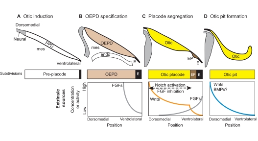
Fig. 2.
Otic induction from the pre-placode. (A) Otic induction requires extrinsic sources of secreted molecules originating from neural tissue, mesenchyme (mes) and pharyngeal endoderm (endo). (B) FGFs act on the pre-placodal domain (PPD) to specify the otic-epibranchial placode domain (OEPD) as separate from ectoderm (E). (C) The OEPD field is further segregated into the otic placode (Otic) and the epibranchial placodes (EP). The otic placode forms under the influence of high Wnts and low FGF signaling. This begins with a Wnt gradient that develops a sharp transition point through feedback loops involving Notch activation and FGF inhibition. (D) By the time the otic pit begins to invaginate, the dorsomedial domain may already be receiving higher Wnt and BMP signals from the adjacent hindbrain, thereby initiating both dorsal-ventral (DV) and mediolateral patterning (not shown). Note that in this and subsequent figures, illustrations of gradients of signaling proteins or their resulting activities are not based on direct observation but rather are speculative, taking into consideration results from experimental embryology, known gene expression patterns and/or phenotypes resulting from perturbations of signaling pathways using drugs or in mutant mice.

