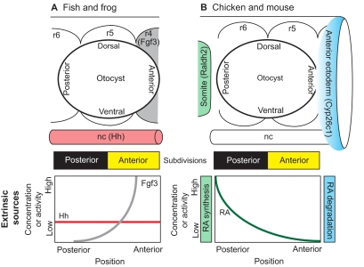
Fig. 4.
Anterior-posterior axial patterning in the inner ear. Extrinsic sources of signaling molecules that pattern the otic placode/vesicle differ between anamniotes and amniotes. (A) Zebrafish and Xenopus otic vesicles begin with a mirror-image symmetric prepattern of otic tissue that is centered along the anterior-posterior (AP) axis adjacent to rhombomere (r) 5. The symmetric template is then independently endowed with posterior identity via hedgehog (Hh) signaling originating from the notochord (nc) and anterior identity via Fgf3 signaling originating from rhombomere 4. (B) In chicken and mouse, the otocyst is centered next to the r5/r6 boundary. AP positional information is influenced by an extrinsic gradient of retinoic acid (RA). This gradient arises because of asymmetry in the synthesis and degradation of RA by enzymes (Raldh2 and Cyp26c1) present in the somatic mesoderm and anterior ectoderm, respectively. There are no lineage data to suggest that the otic field becomes fully subdivided into anterior and posterior halves, as shown schematically by the yellow and black bars for both fish/frog and chicken/mouse. However, the observation of mirror-image symmetry along this axis in mutants or following experimental manipulations supports the idea that there are two distinct compartments.

