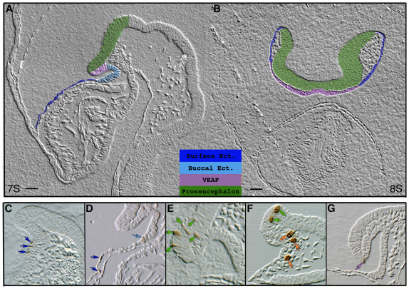
Fig. 2.
Histological sections showing HRP-labelled cells after 1 day of culture. (A,B) Parasagittal (A) and frontal (B) sections of cultured mouse embryos showing painted anatomical domains colonised at E8.5 (7- or 8-somite stage). (C-G) Longitudinal (D,E,G) and frontal (C,F) histological sections with examples of contribution to (C,D) surface ectoderm (dark-blue arrows), (D) buccal ectoderm (light-blue arrow), (E,F) forebrain neuroectoderm (green arrows), (F) emigrating neural crest cells at the level of the forebrain (orange arrows) and (G) ventral ectoderm of the anterior prosencephalon (VEAP; purple arrow). Scale bars: 25 μm in A; 30 μm in B.

