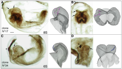
Fig. 7.
Whole-mount images of HRP-labelled mouse embryos and corresponding representation of the clones plotted in the 3D reconstruction. (A,B) Lateral (A) and dorsal (B) views of clone 17 at the 8-somite stage showing two distinct groups of labelled descendant cells (arrows) in the left headfold. Each group presumably derives from one of the documented siblings. (C,D) Lateral (C) and frontal (D) views of clone 34 at the 6-somite stage showing labelled descendants in the neuroectoderm and the VEAP of the left headfold (arrows). Arrowheads mark the descendants of the injected visceral endoderm cell. Scale bar: 130 μm.

