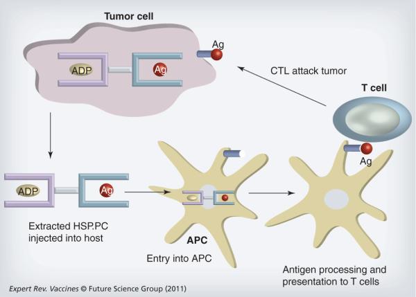
Figure 2. Heat shock protein vaccines prepared from tumor lysates.
Heat shock protein (HSP)A and HSPC family members chaperone intracellular antigens in tumor cells (see Figure 1). When tumor cells are lysed, HSP-peptide complexes (HSP.PC) can be isolated biochemically and form the basis for development of HSP vaccines. The vaccines are then injected into the periphery of tumor bearing hosts where they encounter APCs. APCs are able to take up the HSP.PC and transport them to the sites of antigen processing where the peptide antigens are proteolytically processed, loaded onto MHC class I molecules and presented on the APC surface. APCs (usually dendritic cells [DCs]) then traffic to the efferent lymph nodes where they encounter cognate CD8+ lymphocytes that recognize the tumor antigen-MHC class I complex on the DC surface through their T-cell receptors. T cells then become activated into proliferating CTLs and traffic to the tumor where, under the correct inflammatory circumstances they can extravasate, enter the tumor milieu and kill tumor cells expressing surface MHC class I–antigen complexes with high specificity.
Ag: Antigen; APC: Antigen-presenting cell; CTL: Cytotoxic T lymphocyte.

