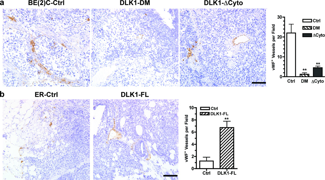
Figure 4. DLK1 enhances angiogenesis in NB xenografts.
Parafin-embedded sections of (a) BE(2)C xenografts (Ctrl, DLK1-DM or DLK1-ΔCyto) or (b) ER xenografts (Ctrl or DLK-FL) were incubated with an anti-von Willebrand Factor (vWF) antibody as described in Materials and Methods. Bar = 100 µm. vWF+ cells were counted from three random fields (mean ± sem, **p<0.001 versus control).

