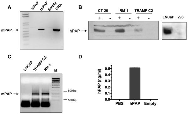
Figure 2. PAP expression.
(A) RT-PCR analysis of Vero cells infected with bPΔ6-mPAP shows mPAP mRNA expression. Cells were grown in 6-well plates and infected at 90-95% confluence with either bPΔ6-hPAP, bPΔ6-mPAP or bPΔ6-empty viruses at MOI=2. After 2 hrs, virus inoculum was removed and replaced with medium DMEM/1% inactivated FCS. Eight hours later the total RNA fraction was isolated from cell lysates using TRIZOL (Invitrogen, USA), and treated with RQ1 RNase-free DNase I (Promega, Madison, WI). Reverse transcription was carried out using SuperScriptIII First Strand Synthesis kit (Invitrogen, USA) and random hexamer primers. cDNAs were subjected to conventional PCR (5 min at 95°C then 28 cycles: 30 seconds at 95°C, 15 seconds at 68°C, 20 seconds at 72°C; and terminal elongation 2 min at 72°C) with specific primers: P1mPAP619 5′ – gcttcctggacaccttgtcgtcgctgtcg – 3′ and P2mPAP1214 5′ – attccgtccttggtggctgc – 3′, designed to generate a PCR product of 595 bp. As a positive control, plasmid DNA (pVec92-mPAP) was used and the PCR reaction was performed without the reverse transcription step. The reaction product was analyzed by 1.2% agarose gel electrophoresis and stained with ethidium bromide. (B) Western blot analysis of hPAP expression in CT-26 colorectal carcinoma 13, RM-1 and TRAMP-C2 cell lines after infection with bPΔ6-hPAP (indicated by +) or bPΔ6-empty (indicated by -) at MOI=2. Human prostate adenocarcinoma LNCaP cells 24 and human embryonic kidney 293 cells were used as controls. Cell lysates were prepared using RIPA buffer 36 24 hours post-infection. Each lysate was separated by SDS-polyacrylamide electrophoresis, transferred to PVDF membrane (BioRad), and immunoblotted with anti-hPAP (DAKO Rabbit anti-Human PAP, A0627) antibody (diluted 1:1000) using standard procedures. (C) Mouse prostate cancer cells express PAP. Total RNA was isolated from LNCaP, TRAMP-C2 and RM-1 cells and RT-PCR was performed using primers previously described 37. mPAP mRNA was observed in both TRAMP-C2 and RM -1 cells but not in human LNCaP prostate cancer cells. (D) In vivo expression of hPAP. TRAMP-C2 cells (5×10⁶/mouse) were implanted subcutaneously into the flanks of male six to eight-week-old C57/BL6 mice (National Cancer Institute, Frederick, MD). Once tumors were established (50-10 mm3) mice were treated intraneoplastically once with bPΔ6-hPAP (2×10⁷ pfu). After 48 hours, tumors were harvested, homogenized and centrifuged. Supernatants were assayed for hPAP using an ELISA Kit (R&D Systems, Minneapolis, MN) according to manufacturer’s instructions. Error bars represent the standard deviation of 3 measurements. All in vivo procedures were approved by the Massachusetts General Hospital Subcommittee on Research Animal Care.

