
Figure 3.
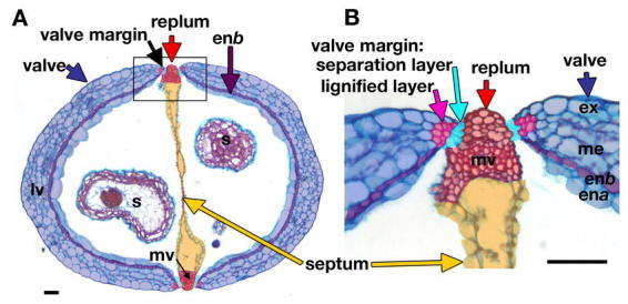
Cross section of a wild-type fruit. (A) Cross section of the ovary of a stage 17 wild-type (Ler) fruit. The fruit has been false colored to distinguish the parts as described in Figure 1. The septum (light orange) divides the fruit into two locules. The valve contains the enb layer (purple), which is lignified. The box encloses the region shown at higher magnification in B. The medial vascular bundle (mv) in the replum and the lateral vascular bundle (lv) in the valve are indicated, as are the developing seeds (s). (B) Close up of a cross section of the replum and valve margin. The valve margin consists of a lignified layer (pink) and the separation layer (aquamarine blue). The valve usually contains 6 cell layers. The outer epidermis is termed the exocarp (ex). The next three cell layers are the mesocarp (me). The inner two cell layers form the endocarp consisting of the lignified enb layer (purple) and the large cells of the ena layer. The scale bars in A and B represent 50 µm.