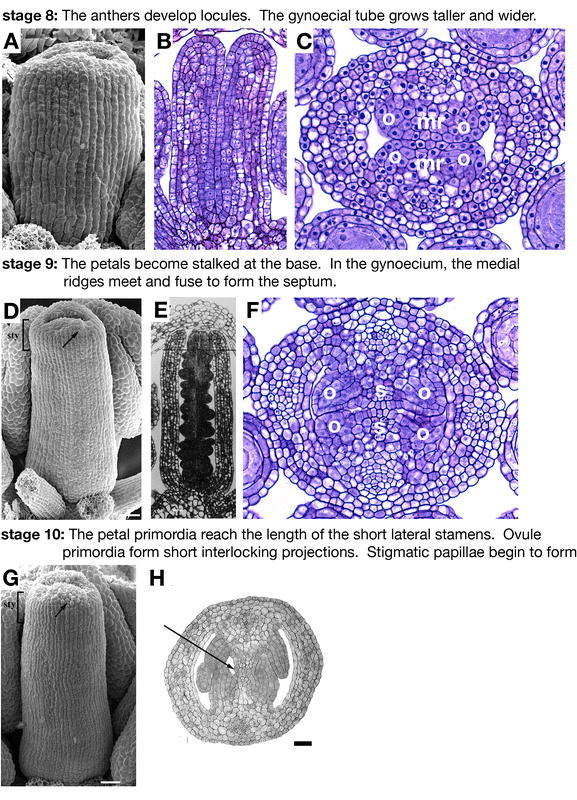
Figure 5.

Flower development: stages 8–10. (A) SEM of a stage 8 gynoecium. (B) Longitudinal section of a stage 8 gynoecium. (C) Transverse section of a late-stage 8 gynoecium. Placentas (o) form on both sides of the medial ridges (mr). (D) SEM of a stage 9 gynoecium. The style region (bracketed) can be identified as morphologically distinct from the ovary. A few of the stigmatic papillae become visible as rounded cells on the top of the gynoecium (arrow). (E) Longitudinal section of a stage 9 gynoecium along the lateral axis. (F) Cross section of a stage 9 gynoecium. The three different classes of cells relating to the future exocarp, mesocarp, and endocarp can be distinguished in the valves. The ovule primordia (op) arise from the placentas flanking the medial ridges. The medial ridges meet in the center of the fruit to form the septum (s). (G) SEM of a stage 10 gynoecium. More stigmatic papillae (arrow) are evident at the top of the gynoecium, which has closed over. (H) Cross section of a stage 10 gynoecium. Transmitting tract precursors (arrow), smaller darkly staining cells, are present in the middle of the septum. Scale bars in D and G represent 25 µm. A–C and F: Reprinted from Current Topics in Developmental Biology, 45, Bowman, J.L., Baum, S.F., Eshed, Y., Putterill, J., and Alvarez, J., Molecular genetics of gynoecium development in Arabidopsis, 155–205, Copyright (1999), with permission from Elsevier. D, G, and H: from Alvarez et al., 2002. E: from Sessions 1997.
