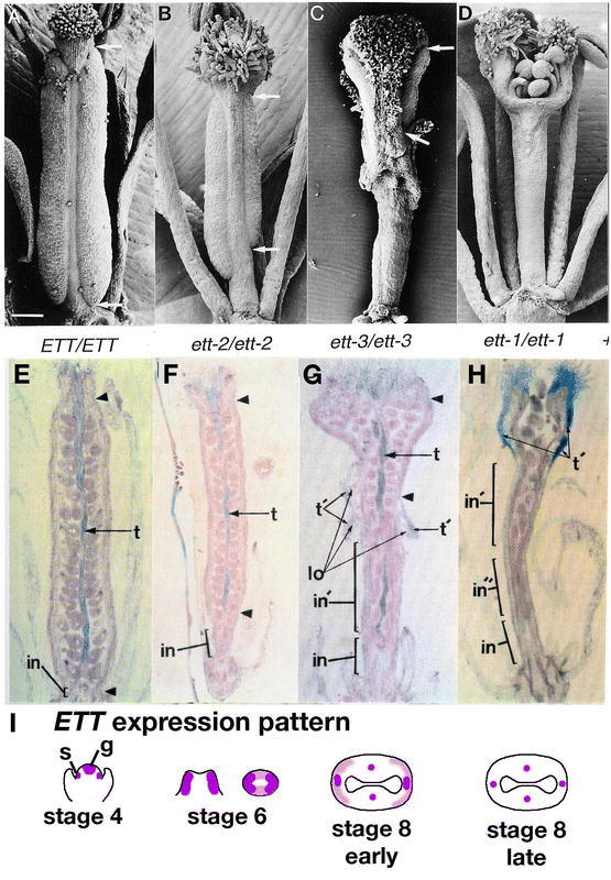
Figure 32.

ETTIN and the apical/basal axis of the gynoecium. (A) Wild-type gynoecium post-pollination. The arrows mark the extent of valve elongation. (B) Gynoecium of the weak ett-2 allele. Note the uneven valves. (C) Gynoecium of the intermediate ett-3 allele. Note the small valves surrounded entirely by stigmatic and stylar tissue. (D) Gynoecium of the strong ett-1 allele. Valves are completely absent. (E) Longitudinal section of a wild-type gynoecium. The blue color indicates transmitting tract cells. t = transmitting tract; t′ = everted transmitting tract; in = internode with a solid stem; in’ = externally gynophore stalk, but contains ovules; in″ = solid gynophore stalk that has formed from a postgenital fusion event; lo = lateral outgrowths. (F) ett-2 with a slightly elongated gynophore (in). (G) ett-3 with some transmitting tract on the outward projections (t′). (H) ett-1 gynoecium with transmitting tract cells primarily on the outer apical surfaces. Abnormal ovules are present on part of the gynophore (in′). (I) Diagram depicting the expression pattern of ETT based on in situ hybridization (Sessions et al., 1997). At stage 4, ETT is expressed in the stamen primordia (s) and the gynoecium primordium (g). At stage 6, ETT is expressed abaxially in the gynoecium. ETT expression is stronger in the lateral regions of the gynoecium than the medial regions. By early stage 8, ETT is strongly expressed in the medial and lateral vascular bundles and weakly expressed abaxially the gynoecium. By late stage 8, ETT expression is only detected within the vascular bundles of the gynoecium. Scale bar in A represents 153 µm. A-H: from Sessions and Zambryski, 1995.
