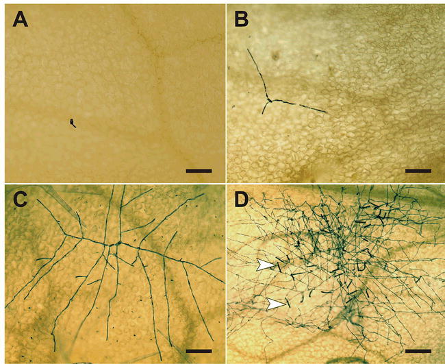
Figure 4.

Microscopic analysis of the development of a powdery mildew colony.
The micrographs show the expansion of a G. orontii colony on the surface of a Col-0 rosette leaf. The series of events starts with a germinated spore at 24 hours post-inoculation (A) (see also Figure 2 for further details on spore germination) and continues with initial hyphal elongation (following successful establishment of the first haustorium inside a host cell) at 48 hours post-inoculation (B). Subsequently, a multi-branched mycelium develops (C; photo taken at 63 hours post-inoculation) and the appearance of numerous conidiophores (arrowheads) from a fully expanded fungal colony from 5 days post-inoculation onwards completes the asexual life cycle (D). Fungal structures were highlighted by Coomassie Blue staining of cleared leaf samples. Scale bar: 100 μm (A–C), 200 μm (D).
