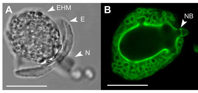
Figure 5.

Haustorial complexes of G. orontii.
Phase contrast (A) and epifluorescence (B) micrographs of G. orontii haustoria isolated from Arabidopsis leaves. Notice the highly convoluted and complex folding of the haustorial cell surface providing a large area for nutrient uptake from and effector delivery into the host. Haustoria were labeled with wheat germ agglutinin-FITC. EHM: extrahaustorial membrane, E: encasement, N: haustorial neck, NB: neck band. Scale bars: 20 μm.
