
Figure 4.
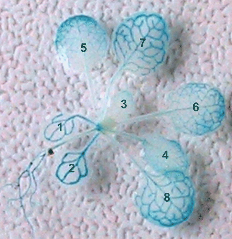
β-Glucuronidase assay of the localization of promoter activity of the source specific AtSUC2, a companion cell specific sucrose transporter. The picture shows promoter activity in vascular bundles of leaves, stems and roots. Cotyledons (leaf No. 1,2) GUS staining follows the transition from sink to source and is strongest in old leaves and increases in young leaves from bottom to top. (Courtesy by W. Schulze)