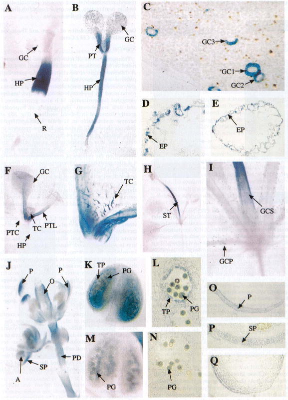
Figure 7.

Staining pattern in transgenic plants containing a CER2-GUS fusion. In Panel A, one-day-old seedlings were germinated on MS medium and stained for GUS activity in the hypocotyl (HP) and guard cells (GC of the leaf). Panel B shows a five day-old seedling with GUS activity in HP, GC and petiole (PT). Panel C is a close-up of the cotyledon from a 5 day-old seedling showing the guard cell staining in the GC of both the adaxial (GC1, GC3) and abaxial (GC2) surfaces of the leaf. Panel D is a cross-section of the hypocotyl of a 5 day-old seedling showing GUS activity mostly in the epidermal layer (EP). Panel E is a cross section of a cotyledonary petiole from a 5 day-old seedling that also shows activity mostly in the EP. Panel F is a 12 day-old plant grown in soil showing GUS activity in the petiole of leaves (PTL), developing trichomes (TC), and GC. Note the lack of activity in the petioles of the cotyledon (PTC) and HP. Panel G is a close-up of the plant in F showing activity in the TC. Panel H is a 22 day-old plant showing strong GUS activity in the stem. Panel I is a close-up view of the plant shown in H that shows strong expression in the guard cells of the petiole (GCP) and stem (GCS). Panel J shows GUS activity in the petals (P), sepals (SP), anthers (A) ovaries (O) and pedicel (PD) of the developing inflorescence. Panel K is a stage 12 anther showing activity in the tapetal layer (TP) and pollen grain (PG). Panel L is a cross-section of a stage 12 anther showing activity in the TP and PG. Panels M and N are a stage 13 anther and cross section showing activity in the PG only. Panels O, P and Q are cross sections of the petal, sepal and ovary showing GUS activity in the EP of petals, across the section in sepals and in the EP of the ovary. Panel R shows activity in the HP of 2 day-old etiolated seedlings but no activity in the root (R). Panel S shows that only cells of the outside of the apical hook (AH) have GUS activity and that the cotyledon (CL) did not stain. Panel T shows staining in a 2 day-old seedling placed in MS medium supplemented with 0.5 mM sodium salicylate for 12 hours. Panel U, shows a 7 day-old seedling transferred to 4 mM BAP in which GUS activity can be seen in all cells of the leaves (TL) grown after BAP induction. Panel V, shows a 7 day-old seedling grown in the absence of BAP shows activity in the petioles and guard cells alone. Panel W is a cross-section of a leaf from a plant grown under the same conditions as the plant in panel U. Note: the staining in all layers differs from that in panel C. Panel X shows a lesser induction of GUS by BAP in a 15 day-old plant grown on BAP-supplemented medium when compared to panel U. Panel Y shows staining in a 10 day-old plant grown for 5 days in MS medium and transferred to MS supplemented with 4 mM BAP for 5 days. In the newly emerging leaves both the trichomes and epidermal cells stain. Taken from Xia et al. (1997).
