
Figure 2.
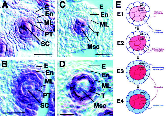
Expression patterns of TPD1 and EMS1/EXS in anthers by in situ hybridization and a model for EMS1/EXS and TPD1 function. (A) and (B) wt anthers at late stage 4. (A) Predominant TPD1 expression in sporogenous cells. (B) Predominant EMS1/EXS expression in the primary tapetum. (C) and (D) wt anthers at early stage 5. (C) TPD1 RNA is detected predominantly in microsporocytes. (D) EMS1/EXS RNA was found mostly in tapetal cells. E, epidermis; En, endothecium; ML, middle layer; Msc, microsporocyte; PT, primary tapetum; SC, sporogenous cell; T, tapetum. Scale bars in (A-D) 20 mm. (E) Model for EMS1/EXS and TPD1 function. (E1) Initially, TPD1 and EMS1 are expressed in the precursors of both meiocytes (the central group) and tapetal cells (the outer ring). (E2) An unknown trigger (?) activates the differentiation of meiocytes, as indicated by shading. (E3) In the differentiating meiocytes, the expression of TPD1 increases and that of EMS1 decreases. The TPD1 protein is secreted and binds to the EMS1 receptors on the neighboring cells, causing an elevation of EMS1 expression in these cells and a drop in TPD1 levels. EMS1 then activates a pathway for tapetal differentiation. (E4) Further reduction of EMS1 and TPD1 in the meiocytes and tapetal cells, respectively, stabilizes the differentiation of tapetal cells. (A-D) From Yang et al., 2003; © 2003 by the American Society of Plant Biologists, used with permission. (E) From Ma, 2005; © 2005 by the Annual Reviews, used with permission.