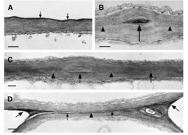
Figure 2.

Ultrastructure of the cuticle of the leaf epidermis of wild type and structural variations of the cuticle in fusion zones between leaves of cutinase-expressing transgenic Arabidopsis plants. (A) Columbia Col-0/gl1. A thin electron-dense cuticle of amorphous structure (arrows) overlays the cell wall polysaccharides in leaves. Bar = 250 nm. (B) and (C) Cutinase-expressing Columbia Col-0/gl1. The fusion zone between leaves is characterized by stretches of a direct contact of the polysaccharides (arrowheads) of the two epidermal cells and the local occurrence of interspersed cuticular material (arrow). Tilting the specimen (C) in the regions of a direct contact of polysaccharides confirmed the absence of detectable amounts of cuticular material or any structural change at the position were both cell walls come into contact (arrowheads). Bars = 250 nm. (D) Cutinase-expressing Columbia Col-0/gl1. The fusion zone is characterized by stretches of low amounts of cuticular material (small arrows) that is partially missing (arrowhead). In contrast to (C), the position where the cell walls of both epidermal cells come into contact is still visible. At the positions of the cell corners where the fusion is tearing apart, electron-opaque polysaccharide material accumulates (large arrows). Bar = 500 nm.
