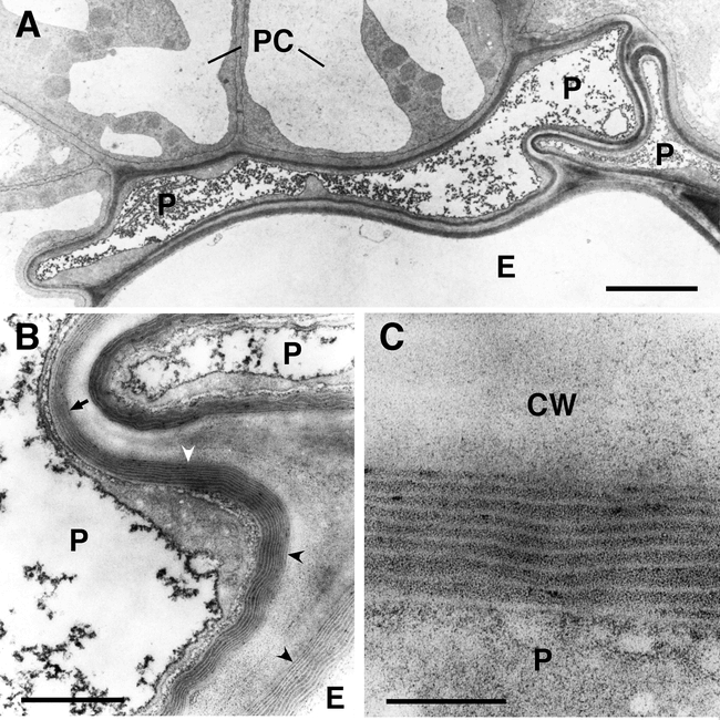
Figure 4.

Ultrastructure of suberized roots tissues of wild type Arabidopsis plants at the beginning of the secondary thickening of the root. (A) Overview of suberized endodermal and peridermal cells in the root. The suberin deposition is visible as electron-opaque layer inside of the primary cell wall. The fully suberized peridermal cell layer typically collapses during the dehydration and embedding procedures necessary for TEM because of the low permeability of the suberized cell walls. Bar: 2.5 micrometers. (B) Enlargement of (A). Fine structure of suberin. The structure of the lamellae with an alternation of electron-opaque and electron-translucent layers of suberin is clearly visible when the specimen is cut perpendicularly to the suberin layers (concave arrowheads). However, the lamellate structure of suberin is barely visible when the specimen in not cut perpendicularly to the suberin layers (arrow). Bar: 500 nm. (C) Enlargement of (B). The thickness of the electron-opaque and electron-translucent layers of the suberin is very regular and characteristic for the tissue sample. Bar: 100 nm. P, peridermal cell; E, endodermal cell; PC, pericycle cell; CW, cell wall.
