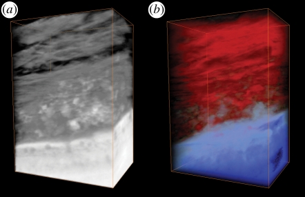
Figure 4.
Three-dimensional reconstructions of the electron tomograms. From the bottom: bulk titanium, surface oxide, apatite layer and bone tissue. The size of the reconstruction is roughly 700 nm high, 550 nm wide and 200 nm thick. (a) The reconstructed area in greyscale. (b) The same region with histogram contrast changes to highlight interior features.

