
Figure 7.
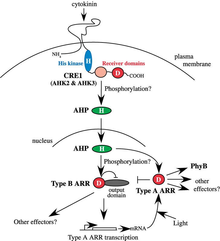
Proposed model for phosphorelay signal transduction in cytokinin signaling.Cytokinin binds to CRE1, and possibly other histidine kinase-like proteins such as AHK2 and AHK3, within the conserved domain flanked by predicted transmembrane domains. CRE1, by analogy to CKI1 (Hwang and Sheen, 2001), is likely to be located in the plasma membrane. Binding of cytokinin activates the transmitter domain (blue), which autophosphorylates on a His (indicated by an H). The phosphate is then transferred an Asp residue (indicated by a D) within the fused receiver domain (red). A second, degenerate receiver domain (pink) is also present. The phosphate is then likely to be transferred to an AHP protein, which translocates to the nucleus where it activates type-B ARRs. The activated type-B ARRs increase the transcription of the type-A ARRs, which feed back to inhibit their own transcription (indicated by ⊥). Light appears to elevate the level of ARR4 protein levels, as indicated by the arrow. The receiver domain of type-B ARRs inhibits the activity of the output domain (gray). The role of phosphorylation of the type-A and type-B ARRs is unclear. The output of this signaling pathway is mostly unknown, although one likely target is PhyB. See text for additional details