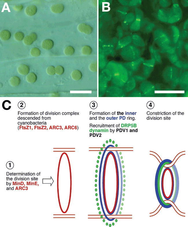
Figure 6.

Plastid division and the division machinery.
(A) Chloroplasts dividing in Arabidopsis hypocotyl cells.
(B) Localization of GFP-tagged DRP5B dynamin protein at the cytosolic side of the division site in Arabidopsis mesophyll cell chloroplasts. (C) Schematic representation of the plastid division machinery. A ‘bacterial’ division complex based on FtsZ forms first at the division site. This is then followed by the formation of the inner and outer PD rings, and finally the recruitment of DRP5B dynamin. Constriction at the vision site then initiates. Scale bars in (A) and (B), 10 μm.
