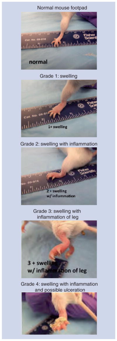
Figure 2. The mouse footpad model of Mycobacterium ulcerans disease.

Grade 1 swelling of the footpad is easily distinguished from the normal footpad 4–8 weeks after infection. Swelling progresses to the grade 2 stage with increasing inflammation. At grade 3, inflammation is apparent further up the leg. If mice are not sacrificed, there can be manifestations of ulceration with cage bedding sticking to the foot. See also Box 1 discussing the model.
