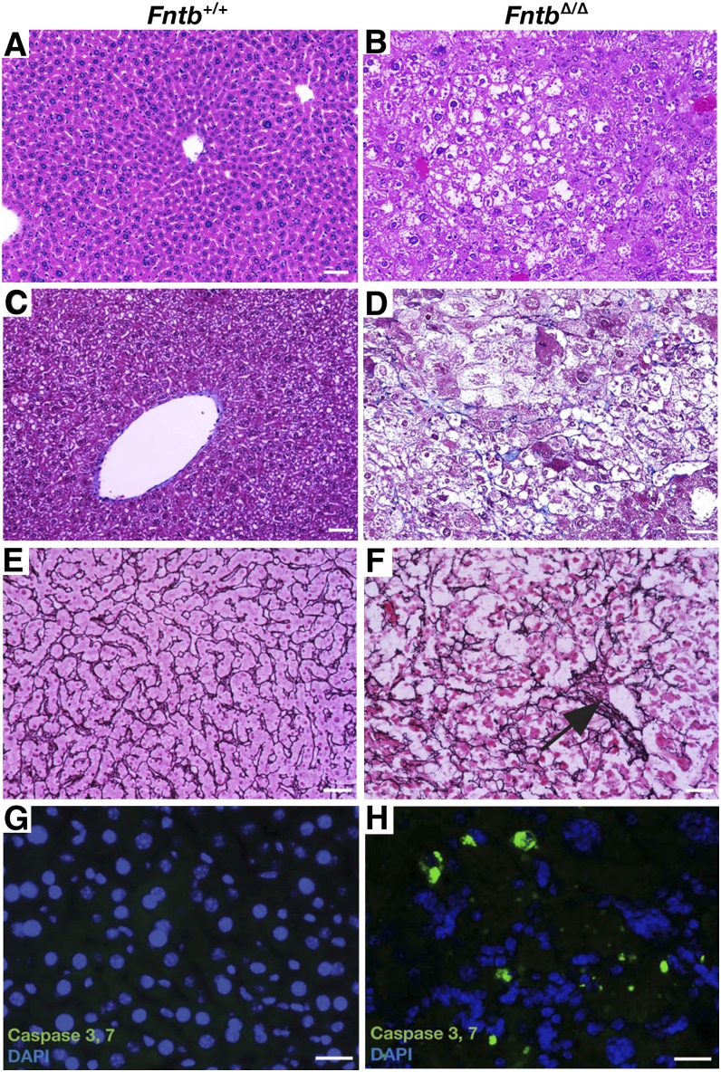
Fig. 2.
Fntb deficiency in the liver causes steatosis, fibrosis, and apoptosis. (A, B) Hematoxylin and eosin–stained sections of liver from 12-month-old Fntb+/+ and FntbΔ/Δ mice, revealing disordered hepatic lobules and steatosis in FntbΔ/Δ mice. (C, D) Masson's trichrome stain revealing increased collagen (blue) in the liver of the FntbΔ/Δ mouse, along with steatosis and ballooning of hepatocytes. (E, F) Reticulin stain (black) revealing a disorganized liver architecture and regions with collapse of reticulin fibers (black arrow), corresponding to areas of hepatocyte loss. Images in A–F were recorded with a 20× objective. Scale bar, 50 µm. (G, H) Fluorescence microscopy revealing active caspases 3 and 7 in the livers of 6-month-old Fntb+/+ and FntbΔ/Δ mice (n = 3 mice/group were examined). Scale bar, 10 µm.

