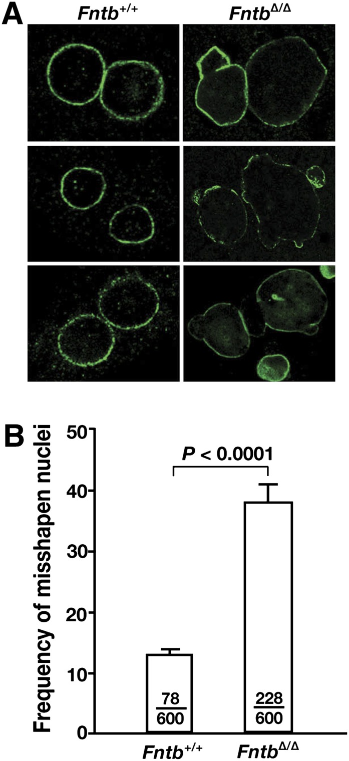
Fig. 4.

Misshapen nuclei in FntbΔ/Δ hepatocytes. (A) Immunohistochemical staining of primary hepatocytes from Fntb+/+ and FntbΔ/Δ mice with an antibody against lamin A (green). (B) Frequency of misshapen nuclei in Fntb+/+ and FntbΔ/Δ hepatocytes. Bars indicate the percentage of cells with misshapen nuclei; the number of cells harboring nuclear blebs and the total number of cells examined are recorded within each bar. Error bars indicate SEM for results with different cell preparations of the same genotype (n = 4/group).
