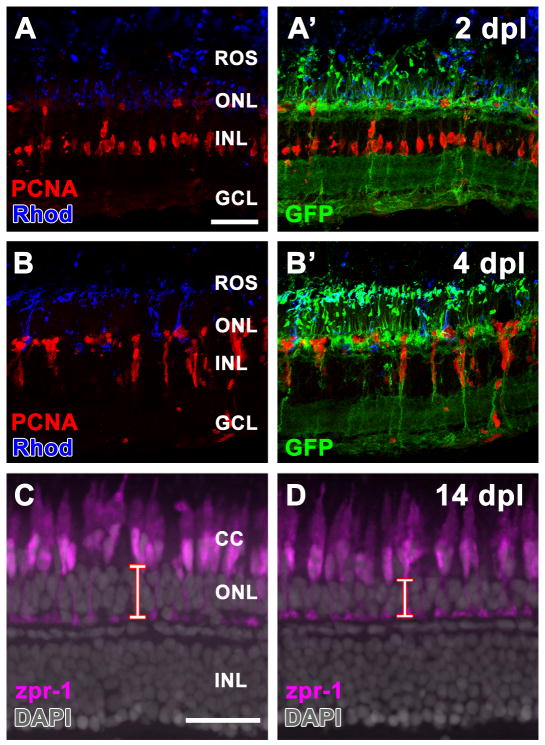
Figure 2. Fgf signaling is required for rod, but not cone, photoreceptor cell regeneration.
A. Retinal section from a Tg(hsp70:dn-fgfr1) adult zebrafish following daily heat shock and 2 days of constant light treatment (2 dpl). Müller glia cell proliferation (PCNA immunolabeling; red) and rod outer segment degeneration (Rhodopsin immunolabeling; blue) are unaffected by loss of Fgf signaling. A'. Overlay of panel A showing immunolocalization of the dn-fgfr1-GFP fusion protein. B. Retinal section from a Tg(hsp70:dn-fgfr1) adult zebrafish following daily heat shock and 4 days of constant light treatment (4 dpl). The continual proliferation of inner nuclear layer progenitors (PCNA immunolabeling; red) and their migration to the outer nuclear layer is unaffected by loss of Fgf signaling. B'. Overlay of panel B showing immunolocalization of the dn-fgfr1-GFP fusion protein. C – D. Retinal sections from wild-type (C) and Tg(hsp70:dn-fgfr1) zebrafish (D) 14 days after intense light damage (14 dpl) and daily heat shock. Double cones are immunolabeled with zpr-1 (magenta). DAPI nuclear stain shows retinal layers. C. Wild-type retinas contain zpr-1+ double cones and thick outer nuclear layer (red bar), which primarily houses rod photoreceptor nuclei. D. The Tg(hsp70:dn-fgfr1) retinas contain zpr-1+ double cones and a thinner than normal outer nuclear layer (red bar). ROS = rod outer segments; CC = cone cells; ONL = outer nuclear layer; INL = inner nuclear layer; GCL = ganglion cell layer. Scale bar: Panel A = 50 μm (A-B'); Panel C = 50 μm (C-D).

