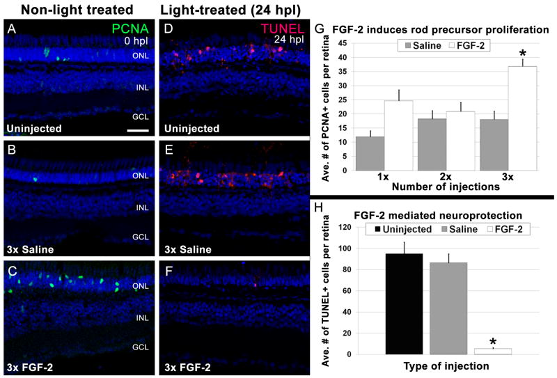
Figure 3. FGF-2 induces rod precursor proliferation and provides neuroprotection.
A - C. Retinal sections from undamaged eyes that were either uninjected (A), or injected three consecutive days with Saline (B) or FGF-2 (C). PCNA immunolocalization (green) shows proliferating cells. Nuclei are stained with TO-PRO-3 (blue). A. A few PCNA-positive rod precursors are present in the outer nuclear layer. B. Saline injections did not significantly increase the number of PCNA-positive rod precursors. C. Large numbers of PCNA-positive rod precursors are observed in the outer nuclear layer in FGF-2-injected retinas. D - F. Retinal sections of 24-hour light-treated zebrafish that were either uninjected (D) or injected three consecutive days with Saline (E) or FGF-2 (F). TUNEL immunolocalization (red) shows cells undergoing apoptosis. Nuclei are stained with TO-PRO-3 (blue). D. Large numbers of TUNEL-positive cells are observed in the outer nuclear layer of uninjected retinas. E. Saline injections did not significantly alter the number of TUNEL-positive nuclei. F. Eyes injected three consecutive days with FGF-2 exhibited significantly fewer TUNEL-positive cells. G. Graphic representation of the average number of PCNA-positive cells in the outer nuclear layer in non-light treated eyes injected with either Saline or FGF-2. H. Graphic representation of the average number of TUNEL-positive cells in 24-hour light-treated retinas that were either uninjected, or injected for three consecutive days with Saline or FGF-2.

