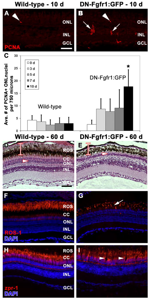
Figure 5. Long-term defect in Fgf signaling results in significant loss of rod, but not cone, photoreceptors.
A. PCNA immunolocalization (red) in a wild-type retina after 10 days of daily heat-shock shows isolated rod precursor cell proliferation in the ONL (arrowhead). B. PCNA immunolocalization (red) in a Tg(hsp70:dn-fgfr1) retina after 10 days of daily heat-shock shows increased proliferation in rod precursor cells (arrowhead; Panel C) and in inner nuclear layer progenitor cells (arrows). C. Graphic depiction comparing the average number of PCNA-positive cells observed in the outer nuclear layer in wild-type and Tg(hsp70:dn-fgfr1) retinas following daily heat-shock over multiple time points. D-I. Wild-type (D, F, H) and Tg(hsp70:dn-fgfr1) (E, G, I) retinas following 60 days of daily heat-shock. D. Histological section of a wild-type retina showing the normal outer nuclear layer (arrowhead) and the thickness of the rod inner and outer segments (bar). E. Histological section of a Tg(hsp70:dn-fgfr1) retina, showing a thin outer nuclear layer (arrowhead) and a near complete loss of rod outer segments (compare the bar in D and E). F. ROS-1 immunolabeling (red) overlaid with DAPI nuclear stain (blue) in a wild-type retina, showing normal rod outer segment thickness and organization. G. A corresponding Tg(hsp70:dn-fgfr1) retinal section, showing ROS-1 immunolocalization (red) to truncated and disorganized rod outer segments (arrow). H. Immunolocalization of zpr-1 to double cones (red) overlaid with DAPI nuclear stain (blue) in a wild-type retina. I. A corresponding Tg(hsp70:dn-fgfr1) retinal section, showing zpr-1 immunolocalization to double cones (red), which are present, but exhibit areas of disorganization (arrowheads). ROS = rod outer segments; CC = cone cells; ONL = outer nuclear layer; INL = inner nuclear layer; GCL = ganglion cell layer. Scale bar: Panel A = 50 μm (A-B'); Panel D = 50 μm (D-I).

