
Figure 6. Same case as in Figure 5 (with 500-μm resolution contrast-enhanced MR angiogram) of left carotid artery confirming intraplaque hemorrhage by black-blood T1-weighted cross-sectional images using 3D magnetization-prepared rapid acquisition gradient echo sequence, where the intraplaque hemorrhage is bright.
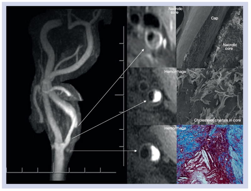
Along the inferior aspect of the intraplaque hemorrhage there is a 1-mm thick fibrous cap between the dark lumen and the bright deep intraplaque hemorrhage. Superiorly there is a well-defined fibrous cap (<500 μm) between the lumen and lipid core. Following endarterectomy, light and scanning electron microscopy demonstrate extensive cholesterol crystals with intraplaque hemorrhage and thin fibrous cap.