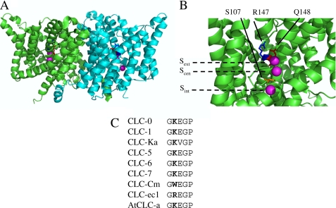
FIGURE 1.
Location of residue Lys210 mapped on the structure of the CLC-ec1 mutant E148Q (PDB entry 1OTU). A, dimeric structure of CLC-ec1 viewed from the membrane plane (extracellular side above and cytoplasmic side below). The two subunits are shown in green and cyan. Residue Gln148 is colored in red, Arg147, corresponding to Lys210 of CLC-5, is shown in blue, and Ser107 in orange. Chloride anions bound to Sext, Scen, Sint are shown in magenta. B, expanded representation of the anion permeation pathway for one of the subunits. The positions of the three binding sites are also indicated by horizontal dashed lines. C, alignment of the sequence stretch comprising Lys210 (shown in bold) for several CLC proteins.

