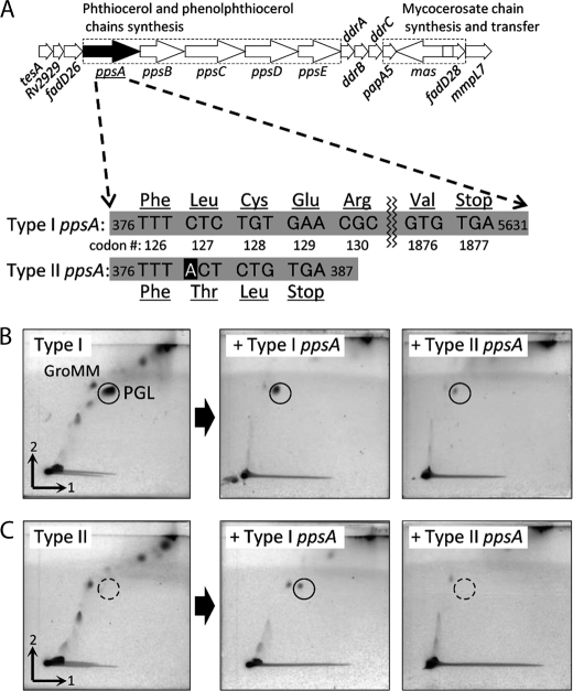
FIGURE 5.
Genetic organization of PGL/PDIM locus (25) and PGL expression. A, the nucleotide sequence of ppsA genes showed that the type II ppsA had a single base insertion and two single base substitutions. The frameshift due to the insertion of this adenine relocated the stop codon from codon 1877 to 129, and an abnormal truncated protein was synthesized. B and C, total lipids extracted from type I (B) and II (C) transformants with type I ppsA or type II ppsA inserted. The manifestation of PGL was confirmed by insertion of the type I ppsA into the type II transformant. The TLC plates were developed with chloroform-methanol (96:4, v/v) first and then toluene-acetone (80:20, v/v). GroMM, glycerol monomycolate.

