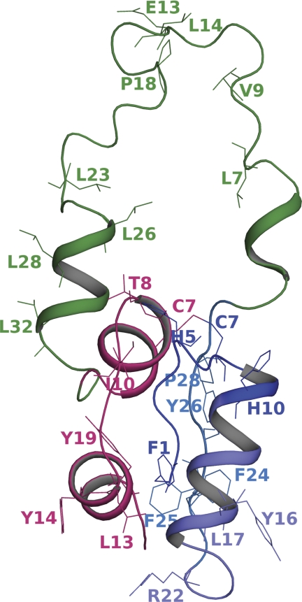
FIGURE 1.
Domain organization of proinsulin and sites of modification. The modeled structure of wild-type proinsulin depicting the amino acid residues undergoing modification in protein footprinting experiments is shown. The A-chain is in magenta, and the C-chain is in green. The three peptide fragments in the B-chain are rendered in different shades of blue.

