Fig. 10.

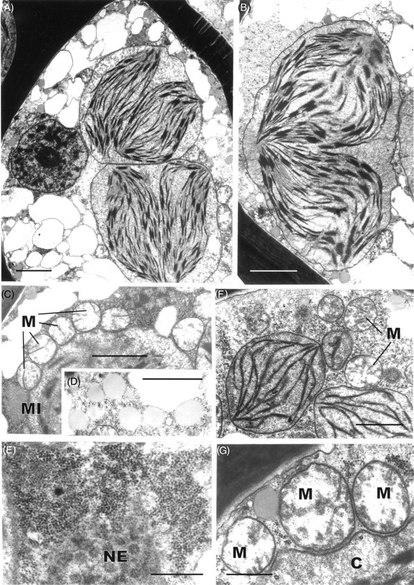
Transmission electron microscopy of dehydrated leaf-lamella (A–E) and lamina (F) cells of Polytrichum formosum. (A) General aspect. Note the condensed chromatin in the nucleus, numerous small vacuoles and two ovoid chloroplasts each containing two groups of thylakoids closely juxtaposed. (B) Detail of ovoid chloroplast containing two groups of thylakoids. (C) Mitochondria with clear stroma and tubular cristae lying close to a chloroplast. (D) Spherical lipid droplets. (E) Aggregation of ribosomes adjacent to the nuclear envelope containing numerous pores. (F) Ovoid chloroplasts in a leaf lamina cell lacking peripheral reticulum. (G) Detail of mitochondria lying adjacent to a chloroplast. C, Chloroplast; M, mitochondrion; MI, microbody; NE, nuclear envelope. Scale bars: A, B = 2 µm; C, D, F = 1 µm; E, G = 0·5 µm.
