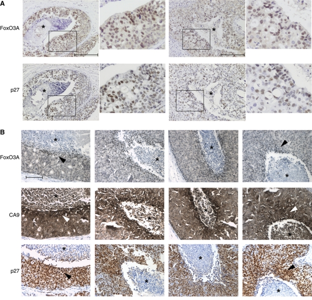
Figure 7.
FoxO3A is activated in hypoxic human tumour tissue. (A) Human samples of carcinoma in situ of the breast were immunohistochemically stained using a FoxO3A antibody. Two different tumours are shown with a magnified view to the right of each. Scale bars indicate 100 μM. (B) Human samples for carcinoma in situ of the breast were analysed for the immunohistochemical localization of FoxO3A, CA9, and p27 in adjacent slides. The necrotic cores of the tumours are highlighted by an *. Scale bars indicate 100 μm.

