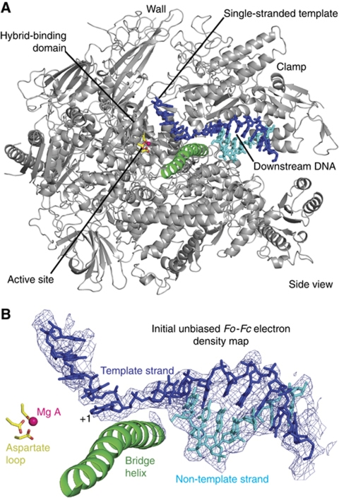
Figure 2.
Structure of a Pol II–DNA complex mimicking part of the OC. (A) Overview. A ribbon model of Pol II is shown in silver, and nucleic acids are shown as stick models. The bridge helix is highlighted in green, the active site metal ion A is in pink, and aspartate side chains holding the metal are in yellow. The view is from the side. (B) Unbiased Fo−Fc difference electron density for DNA contoured at 3σ.

