Figure 1.
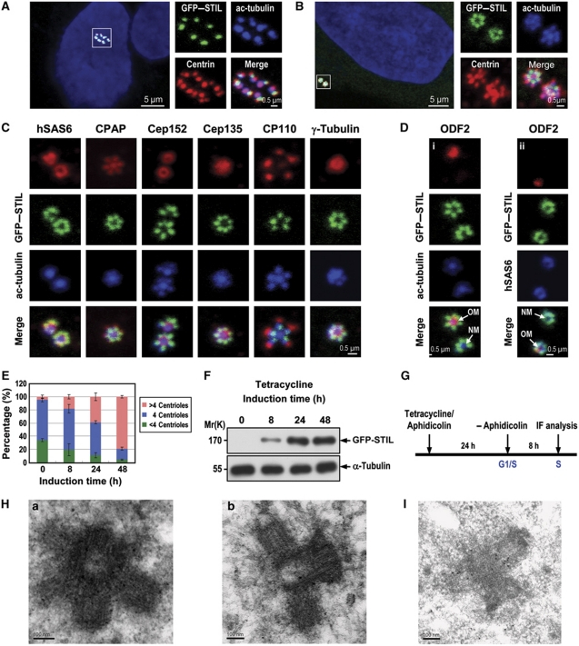
Excess STIL induces centriole amplification. Unsynchronized (A) or synchronized (B–D) U2OS-based GFP–STIL-inducible cells treated as shown in (G) were analysed by confocal fluorescence microscopy using indicated antibodies. Procentriole formation was visualized by anti-centrin and anti-ac-tubulin staining (A, B). GFP–STIL was directly visualized by confocal fluorescent microscopy. DNA was counterstained with DAPI. (C, D) Normal incorporation of several centrosomal proteins into GFP–STIL-induced centrioles. (E) The percentage of amplified centrioles induced by excess GFP–STIL at different time points. Error bars represent mean±s.d. of 200 cells from three independent experiments. (F) Immunoblot analysis of the expression levels of exogenously expressed GFP–STIL at indicated times after tetracycline induction. α-Tubulin was used as a loading control. (G) A protocol to analyse GFP–STIL-induced centriole amplification. (H, I) GFP–STIL-inducible cells were treated as described in (G). Four hours after aphidicolin release, the cells were processed for electron microscopy (H-a, transverse section; H-b, longitudinal section) and immunogold EM (I). OM, old mother centriole; NM, new mother centriole.

