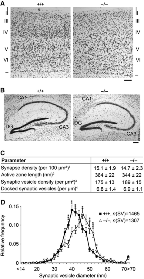
Figure 2.
Significantly increased SV diameters in hippocampal syndapin I KO synapses. (A, B) Histological analyses of cortices (A) and hippocampi (B) of adult WT and syndapin I KO mice via Nissl staining showed no gross defects in brain development and architecture. Borders and the six layers (I–VI) of the cortex are marked (A). DG, dentate gyrus. Bars, 200 μm. (C) Detailed morphometric analyses of synaptic terminals. 1Determined as postsynaptic density (PSD) number from randomly selected electron micrographs (total area of 49.1 μm2 each, n=211 (+/+) and n=259 (−/−)), 2determined as extension of the PSD, 3determined as SV number inside a semicircle around the active zone and 4SVs located ⩽50 nm from the plasma membrane. Parameters 2–4 and SV size distribution (D) were determined from high-magnification micrographs (n=40 (+/+) and n=41 (−/−); 4 animals/genotype). (D) SV size distribution in WT and syndapin I KO animals. *P<0.05; **P<0.01; ***P<0.001.

