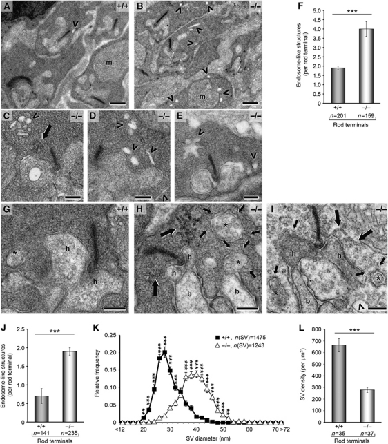
Figure 4.
Drastic alterations in rod photoreceptor ribbon synapses of syndapin I KO animals after light exposure. (A–F) Rod ribbon synapses after acute exposure to light from WT (A) and syndapin I KO (B–E) mice. Note the high number of endosome-like structures (arrowheads) and the occurrence of tubular structures (C, large arrow), respectively, in syndapin I KO synapses. m, mitochondrion. (F) Endosome-like structures were increased by 110% in syndapin I KO mice (n=4 animals/genotype). (G–L) Syndapin I KO synapses (H, I) differ from WT (G) after long exposure to light and showed massive membrane trafficking defects with numerous coated omega-shaped profiles at the presynaptic plasma membrane (H, I, small arrows). In addition, branched tubular structures (H, I, large arrows) and endosome-like structures (I, arrowhead) were visible in syndapin I KO synapses. h, Horizontal cell processes; b, bipolar cell dendrites; *, peripheral protrusions of horizontal cells. (J) Quantification of endosome-like structures per rod terminal from 4 animals/genotype after long exposure to light revealed a 170%-increase. (K) SV sizes were shifted to larger SV diameters. (L) SV density was reduced by 58% in syndapin I KO mice. Quantitative data represent mean±s.e.m.; **P<0.01 ***P<0.001. Scale bars, 0.5 μm (A, B) and 0.2 μm (C–E, G–I).

