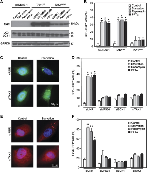
Figure 2.
Reduced interaction between Beclin 1, TAB and TAB3 in conditions of autophagy induction. (A, B) Inhibition of autophagy by dominant-negative (DN) TAK1. HeLa cells were co-transfected with a GFP–LC3-encoding construct plus pcDNA3.1 (empty vector), or plasmids for the expression of WT TAK1 (TAK1WT) or the DN TAK1K63W mutant. One day later, cells were either left untreated (control) or driven into autophagy by starvation or by the administration of 1 μM rapamycin or 30 μM pifithrin α (PFTα), followed by immunoblotting for the detection of TAK1 and endogenous LC3 (A) or immunofluorescence microscopy for the quantification of cells with cytosolic GFP–LC3 puncta (GFP–LC3VAC cells) (B) (mean values±s.d., n=3; *P<0.01 versus control cells). GAPDH levels were monitored to ensure equal loading. (C, D) Inhibition of autophagy by knockdown of VPS34, Beclin 1 (BCN1) and TAK1. siRNAs that effectively deplete VPS34, BCN1 and TAK1 were co-transfected with a GFP–LC3-encoding plasmid in HeLa cells. Autophagy was then induced as in (A) and the frequency of GFP–LC3VAC cells (mean values±s.d., n=3; *P<0.01 versus control cells) was determined. (E, F) The same setting shown in (C, D) was performed with U2OS cells and FYVE–RFP (mean values±s.d., n=3; *P<0.01, **P<0.001 versus control cells).

