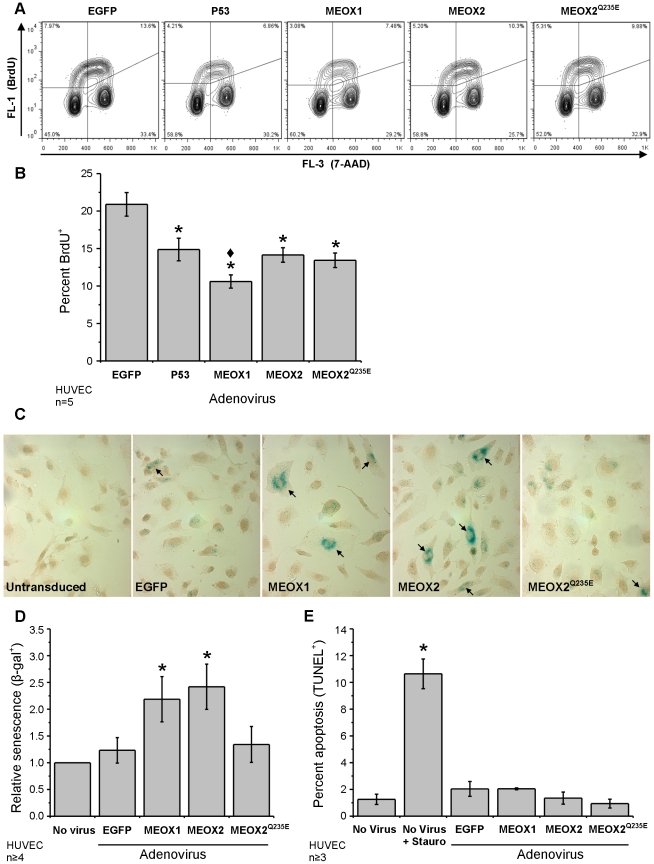
Figure 5. Increased MEOX1 or MEOX2 expression leads to increased endothelial cell senescence.
A) Representative flow cytometry showing the density of BrdU+ endothelial cells (upper left and right quadrants). HUVECs were transduced with N-terminal FLAG tagged MEOX1 and MEOX2 adenoviral constructs at a multiplicity of infection (MOI) of 100; 48 hours later, cells were labeled with BrdU for one hour prior to fixation. DNA was stained with 7-aminoactinomycin D (7-AAD). B) Quantification of the flow cytometry data. Expression of MEOX1, MEOX2 and MEOX2Q235E mutant decreased cellular proliferation comparable to p53 (positive control) as assessed by BrdU incorporation into cycling cells. C) Representative images showing SA-β-gal+ cells (blue). Nuclei were stained with hematoxylin (brown). D) Quantification of SA-β-gal+ cells shows that both MEOX1 and MEOX2 expression increased the number of senescent HUVECs. In contrast, MEOX2Q235E expression did not alter the level of endothelial cell senescence. HUVECs were transduced with N-terminal FLAG tagged MEOX1 and MEOX2 adenoviral constructs at a MOI of 250; 48 hours later cells were fixed and stained. D) MEOX proteins do not induced endothelial cell apoptosis. HUVECs were transduced with FLAG tagged MEOX1, MEOX2 and MEOX2Q235E adenoviral constructs at a MOI of 250; 48 hours later cells were fixed and stained. Staurosporine was used as a positive control for apoptosis induction. * Indicates a statistically significant change (p<0.05) when compared to the EGFP control. ♦ Indicates a statistically significant difference (p<0.05) between MEOX1 and MEOX2.

